Image | Statistics | Caption |
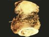 |  | Gross appearance of the specimen after radical hysterectomy: large bulging tumour of the uterine cervix. |
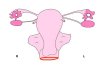
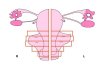
 | | Gross examination of the specimen after radical hysterectomy (scheme). |
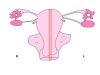
 | | Sampling of the radical hysterectomy specimen - Step 1: section of the vaginal cuff (in red). |
 | | Sampling of the radical hysterectomy specimen - Step 1: section of the vaginal cuff. |
 | | Radical hysterectomy for invasive squamous cell carcinoma: vaginal section showing tumour involvment of the resection margin (arrows). |
 | | Sampling of the radical hysterectomy specimen - Step 2: Longitudinal section of the uterus. |
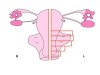 | | Sampling of the radical hysterectomy specimen - Step 3a: transverse sections of the cervical body and parametrium (left). |
 | | Sampling of the radical hysterectomy specimen - Step 3b: sections of the cervical body and parametrium (right). |
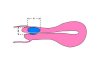 | | Sampling of the radical hysterectomy specimen - Gross section in the sagittal plane to determine the length (or height) (A) and depth (B) of the tumour. |
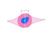 | | Sampling of the radical hysterectomy specimen - Section in the transverse plane to determine the width (C) of the tumour. The tumoral volume = A x B x C (length x depth x width) |
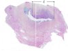 | | Radical hysterectomy - Large histological section in the transverse plane to determine the width (C) of the tumour. |


